Beranda
/ Tendon Diagram : 13 114 Tendon Stock Photos Pictures Royalty Free Images Istock : Biceps tendons the biceps muscle has two tendons at the shoulder, called the long head and short head.
Tendon Diagram : 13 114 Tendon Stock Photos Pictures Royalty Free Images Istock : Biceps tendons the biceps muscle has two tendons at the shoulder, called the long head and short head.
Insurance Gas/Electricity Loans Mortgage Attorney Lawyer Donate Conference Call Degree Credit Treatment Software Classes Recovery Trading Rehab Hosting Transfer Cord Blood Claim compensation mesothelioma mesothelioma attorney Houston car accident lawyer moreno valley can you sue a doctor for wrong diagnosis doctorate in security top online doctoral programs in business educational leadership doctoral programs online car accident doctor atlanta car accident doctor atlanta accident attorney rancho Cucamonga truck accident attorney san Antonio ONLINE BUSINESS DEGREE PROGRAMS ACCREDITED online accredited psychology degree masters degree in human resources online public administration masters degree online bitcoin merchant account bitcoin merchant services compare car insurance auto insurance troy mi seo explanation digital marketing degree floridaseo company fitness showrooms stamfordct how to work more efficiently seowordpress tips meaning of seo what is an seo what does an seo do what seo stands for best seotips google seo advice seo steps, The secure cloud-based platform for smart service delivery. Safelink is used by legal, professional and financial services to protect sensitive information, accelerate business processes and increase productivity. Use Safelink to collaborate securely with clients, colleagues and external parties. Safelink has a menu of workspace types with advanced features for dispute resolution, running deals and customised client portal creation. All data is encrypted (at rest and in transit and you retain your own encryption keys. Our titan security framework ensures your data is secure and you even have the option to choose your own data location from Channel Islands, London (UK), Dublin (EU), Australia.
Tendon Diagram : 13 114 Tendon Stock Photos Pictures Royalty Free Images Istock : Biceps tendons the biceps muscle has two tendons at the shoulder, called the long head and short head.. We are pleased to provide you with the picture named right arm muscle and tendon anatomy.we hope this picture right arm muscle and tendon anatomy can help you study and research. Cook and purdum have proposed a new strategy when approaching tendon pain, and this is called the tendon continuum. For more anatomy content please follow us and visit our website: The foot diagram has a complex structure made up of bones, ligaments, muscles, and tendons.understanding the structure of the foot is best done by looking at a foot diagram where the anatomy has been labeled. Fall on one point of shoulder and can rupture these ligaments with dislocation of ac joint.
Anatomynote.com found right arm muscle and tendon anatomy from plenty of anatomical pictures on the internet. Neck muscle anatomy mri 12 photos of the neck muscle anatomy mri neck muscle anatomy images, neck muscle anatomy pictures, neck muscle anatomy posterior, neck muscle anatomy ultrasound, neck muscles anatomy radiology, human muscles, neck muscle anatomy images, neck muscle anatomy pictures, neck muscle anatomy. Here you can see the tendons that extend down the top of your foot toward your toes, allowing you to curl your toes upward if need be. They suggest that the tendon can move up and down this. Anatomy muscular system hand palm muscle stock vector.
Basic Hand And Wrist Anatomy Hand Institute Of Charleston from handinstituteofcharleston.com The tendon that attaches the biceps muscle to the forearm bones (radius and ulna) is called the distal biceps tendon. The tendons have 2 functions: Back muscle diagrams labeled 12 photos of the back muscle diagrams labeled back muscle diagrams labeled, lower back muscle diagrams labeled, human muscles, back muscle diagrams labeled, lower back muscle diagrams labeled. For more anatomy content please follow us and visit our website: The patellar tendon holds the patella with other two bones, similarly iliotibial band helps in supporting tibia and fibula. Tendons, located at each end of a muscle, attach muscle to bone. Learn about the anatomy and physiology of tendons. The achilles tendon attaches the muscles of the calves to the bones of the ankle and foot.
The long head of biceps (lhb) is a very important tendon that travels through the shoulder joint (glenohumeral joint).the biceps tendon begins at the top of the shoulder socket (the glenoid) and then passes across the front of the shoulder to connect to the biceps muscle.
Your musculoskeletal system includes bones, muscles, tendons, ligaments and soft tissues. The tendon runs down the back of your lower leg from the back of the knee to the heel. Biceps tendons the biceps muscle has two tendons at the shoulder, called the long head and short head. The achilles tendon is also called the calcaneal tendon. The muscles, bones, ligaments, and tendons in the back can all be injured and cause back pain. Tendons, located at each end of a muscle, attach muscle to bone. The tendons have 2 functions: Related posts of shoulder muscles and tendons diagram neck muscle anatomy mri. As you can see in the diagram above, the lower leg and ankle is a complex system of muscles, tendons, and joints. Anatomynote.com found right arm muscle and tendon anatomy from plenty of anatomical pictures on the internet. Cook and purdum have proposed a new strategy when approaching tendon pain, and this is called the tendon continuum. The patellar tendon connects the apex of the patella to the tibial tuberosity, and improves the way the quadriceps muscle pulls on the tibia. The largest structure in the above schematic is the tendon (shown) or the ligament itselt.
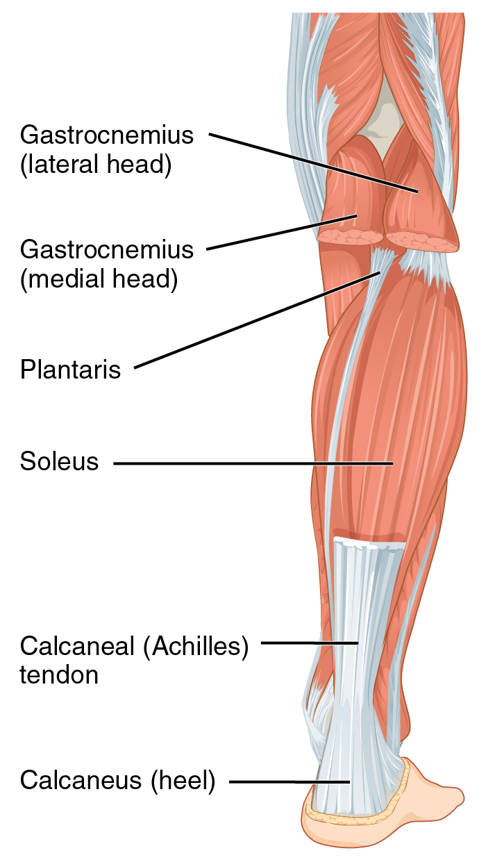
You can see a diagram of the achilles tendon below. Tendons in the knee play a very important role in holding the knee and the muscles together. Allows the action of raising the foot. Diagram of the ankle bones. The bones of the hip include the femur, the ilium, the ischium, and the pubis.
Achilles Tendon Wikipedia from upload.wikimedia.org The reactive tendinopathy, tendon disrepair and the degenerative tendinopathy. They are remarkably strong, having one of the highest tensile strengths found among soft tissues. We are pleased to provide you with the picture named right arm muscle and tendon anatomy.we hope this picture right arm muscle and tendon anatomy can help you study and research. Also allows the action of raising up onto toes. Related posts of shoulder muscles and tendons diagram neck muscle anatomy mri. Back muscle diagrams labeled 12 photos of the back muscle diagrams labeled back muscle diagrams labeled, lower back muscle diagrams labeled, human muscles, back muscle diagrams labeled, lower back muscle diagrams labeled. Tendons are found throughout the body, from the head and neck all the way down to the feet. The long head of biceps (lhb) is a very important tendon that travels through the shoulder joint (glenohumeral joint).the biceps tendon begins at the top of the shoulder socket (the glenoid) and then passes across the front of the shoulder to connect to the biceps muscle.
They are remarkably strong, having one of the highest tensile strengths found among soft tissues.
Brings trunk forward, and aids expiration. Tendons are found throughout the body, from the head and neck all the way down to the feet. The achilles tendon is also called the calcaneal tendon. Your musculoskeletal system includes bones, muscles, tendons, ligaments and soft tissues. The ligament or tendon then is split into smaller entities called fascicles. Diagram of knee tendons and ligaments. Tendons in the knee play a very important role in holding the knee and the muscles together. If you would like to learn all the parts of the foot structure, you have come to the right place. They suggest that the tendon can move up and down this. The achilles tendon is the largest. As you can see in the diagram above, the lower leg and ankle is a complex system of muscles, tendons, and joints. The largest structure in the above schematic is the tendon (shown) or the ligament itselt. The foot diagram has a complex structure made up of bones, ligaments, muscles, and tendons.understanding the structure of the foot is best done by looking at a foot diagram where the anatomy has been labeled.
The tendon that attaches the biceps muscle to the forearm bones (radius and ulna) is called the distal biceps tendon. The fascicle contains the basic fibril of the ligament or tendon, and the fibroblasts, which are the biological cells that produce the ligament or tendon. We are pleased to provide you with the picture named right arm muscle and tendon anatomy.we hope this picture right arm muscle and tendon anatomy can help you study and research. Ligaments and tendons serve similar purposes, but in different ways. Attaches the calf muscles to the calcaneus, most important muscles for running, jumping, walking etc.
Extensor Muscle Anatomy Britannica from cdn.britannica.com They propose there are 3 stages to this continuum. The bones of the hip include the femur, the ilium, the ischium, and the pubis. Diagram of knee tendons and ligaments. Hip, thigh, leg & tendon muscle diagrams. The patellar tendon holds the patella with other two bones, similarly iliotibial band helps in supporting tibia and fibula. Tendons are the connective tissues between the bones and the muscles. For more anatomy content please follow us and visit our website: This important tendon in the back of the calf and ankle stores the elastic energy needed for running, jumping, and other physical activity.
They propose there are 3 stages to this continuum.
This important tendon in the back of the calf and ankle stores the elastic energy needed for running, jumping, and other physical activity. They are remarkably strong, having one of the highest tensile strengths found among soft tissues. It can be used by a teacher or student for academic purposes. Diagram of inside the body. The fascicle contains the basic fibril of the ligament or tendon, and the fibroblasts, which are the biological cells that produce the ligament or tendon. One tendons inserts onto the forearm bone, the radius, and the second spreads out to join the fascia along the upper part of the forearm. The long head of biceps (lhb) is a very important tendon that travels through the shoulder joint (glenohumeral joint).the biceps tendon begins at the top of the shoulder socket (the glenoid) and then passes across the front of the shoulder to connect to the biceps muscle. The reactive tendinopathy, tendon disrepair and the degenerative tendinopathy. 2 ligaments (trapezoid& conoid ligaments) attach the clavicle coracoid process of scapula these tiny ligaments (w/ acominoclavicular joint) keep scapula attached to clavicle. Fall on one point of shoulder and can rupture these ligaments with dislocation of ac joint. Tendons, located at each end of a muscle, attach muscle to bone. Brings trunk forward, and aids expiration. Tendons that make this possible include: